Chest Muscle Anatomy Diagram : Upper extremity - Occupational Therapy 205 with Teresa at Tufts University - StudyBlue. They are categorized by the muscles which they affect (primary and secondary), as well as the equipment required. Meet your pectoralis major and pectoralis minor. Typically, one attachment remains stationary and is called the origin and the other attachment moves. #chest muscle anatomy and exercises #chest muscle chart #chest muscle diagram workout #muscle diagram of chest #muscle diagram of the chest. The chest anatomy includes the pectoralis major, pectoralis minor and the serratus anterior.
Surrounding the rotator cuff muscles are many groups of muscles that work together to produce the various movements of the shoulder. 367 x 280 jpeg 23 кб. Anatomy • free medical books. Typically, one attachment remains stationary and is called the origin and the other attachment moves. Female chest muscle anatomy diagram ~ diagram.
Muscle Anatomy Workout Image from weighteasyloss.com Chest anatomy images, stock photos & vectors | shutterstock. You may also find triceps, lateral head brachialis anatomynote.com found chest muscle anatomy from plenty of anatomical pictures on the internet. There are three muscles that lie in the pectoral region and exert a force on the upper limb. The movement that results from contraction is called the action of the muscle. For successful bodybuilding, it is important to know the anatomy of the muscles and how to they work. #chest muscle anatomy and exercises #chest muscle chart #chest muscle diagram workout #muscle diagram of chest #muscle diagram of the chest. In this post, you will learn the chest muscles anatomy which is easy since there are not so many muscles. The dominant muscle in the upper chest is the pectoralis major.
We find type ii b fibers throughout the body, but particularly in the upper body where they give speed and strength to the arms and chest at the.
Download human muscle anatomy diagram vector art. 1300 x 1390 jpeg 297 кб. See more ideas about muscle diagram, medical anatomy, muscle anatomy. A massive chest anchors the upper body and enhances the. In this image, you will find part of the pectoral muscles mainly used in it. Want to learn more about it? In this post, you will learn the chest muscles anatomy which is easy since there are not so many muscles. Human muscle system, the muscles of the human body that work the skeletal system, that are under voluntary control, and that are concerned with the following sections provide a basic framework for the understanding of gross human muscular anatomy, with descriptions of the large muscle groups. Typically, one attachment remains stationary and is called the origin and the other attachment moves. The chest anatomy includes the pectoralis major, pectoralis minor and the serratus anterior. Find out more about the individual muscles within the chest anatomy by clicking their respective links throughout this page. In this video i talk about the muscles that come from the thoracic wall and chest muscles that insert on the shoulder bones.✅. The two sides connect at the sternum, or breastbone.
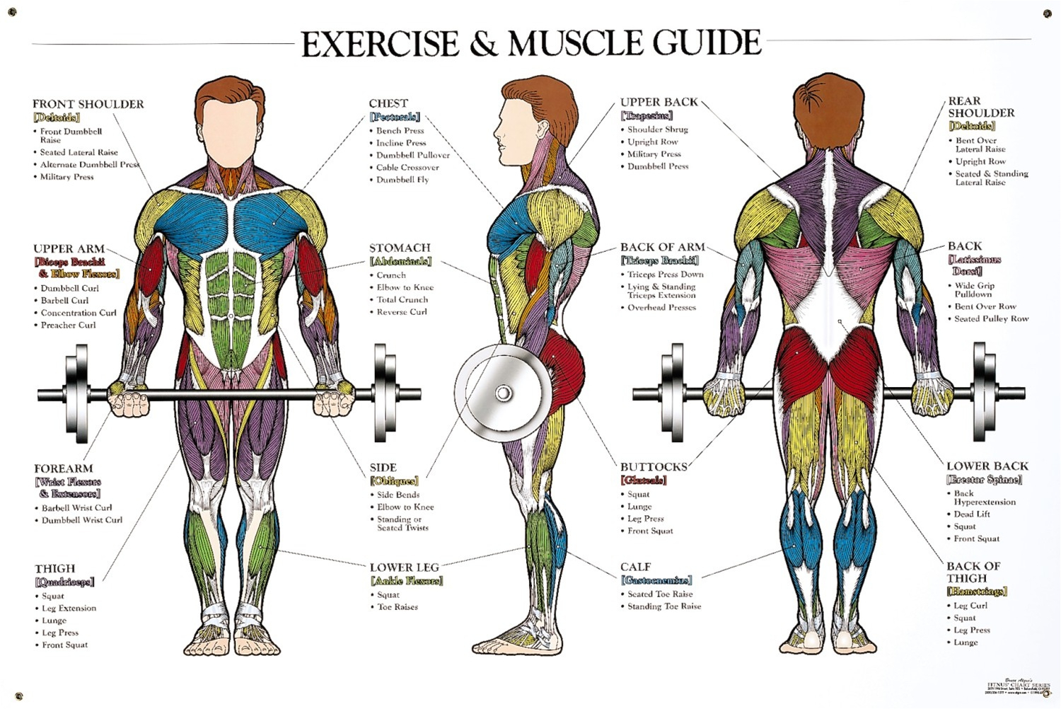
There are three muscles that lie in the pectoral region and exert a force on the upper limb. Anatomy of the chest and the lungs: The major muscle in the chest is the pectoralis major. Want to learn more about it? This page provides an overview of the chest muscle group.
Muscles of Face, Neck, Chest & Abdomen - Kinesiology 270 with Gordon at University of ... from classconnection.s3.amazonaws.com We think this is the most useful anatomy picture that. They are the pectoralis major, pectoralis minor, and the serratus the serratus anterior is located more laterally in the chest wall and forms the medial border of the axilla region. Learn about anatomy diagram muscle with free interactive flashcards. Note that the middle lobe bronchi are relatively anterior (right. Almost all muscles cross at least one joint (moveable connection between two bones) and cause an action across that joint. The two sides connect at the sternum, or breastbone. Choose from over a million free vectors, clipart graphics, vector art images, design templates, and illustrations created by artists worldwide! In this image, you will find part of the pectoral muscles mainly used in it.
Find out more about the individual muscles within the chest anatomy by clicking their respective links throughout this page.
Almost all muscles cross at least one joint (moveable connection between two bones) and cause an action across that joint. Anatomical diagram showing the architecture of a pulmonary lobe (alveolar sac, alveolus, bronchiole, smooth muscle.) Each organ or muscle consists of skeletal muscle tissue, connective tissue, nerve they range from extremely tiny strands such as the stapedium muscle of the middle ear to large masses such as the muscles of the thigh. We think this is the most useful anatomy picture that. #chest muscle anatomy and exercises #chest muscle chart #chest muscle diagram workout #muscle diagram of chest #muscle diagram of the chest. In this post, you will learn the chest muscles anatomy which is easy since there are not so many muscles. 367 x 280 jpeg 23 кб. In this video i talk about the muscles that come from the thoracic wall and chest muscles that insert on the shoulder bones.✅. Note that the middle lobe bronchi are relatively anterior (right. Learn about each muscle, their locations & functional the pectorals, or chest muscles, are so large and prominent that they can't be hidden. Meet your pectoralis major and pectoralis minor. 1300 x 1390 jpeg 297 кб. See more ideas about muscle diagram, medical anatomy, muscle anatomy.
O muscles—sternocleidomastoid, anterior and middle scalene, infrahyoid, pectoralis major and minor, deltoid, trapezius, infraspinatus, supraspinatus, subscapularis diagrams of normal airway anatomy, lateral views. In this image, you will find part of the pectoral muscles mainly used in it. Female chest muscle anatomy diagram ~ diagram. 367 x 280 jpeg 23 кб. The dominant muscle in the upper chest is the pectoralis major.
Musculature Anatomy Chart In Color | Musculature anatomy chart / Musculature anatomy chart ... from i.pinimg.com Surrounding the rotator cuff muscles are many groups of muscles that work together to produce the various movements of the shoulder. Almost all muscles cross at least one joint (moveable connection between two bones) and cause an action across that joint. Human muscle system, the muscles of the human body that work the skeletal system, that are under voluntary control, and that are concerned with movement, posture, and balance. A whole skeletal muscle is considered an organ of the muscular system. In this image, you will find part of the pectoral muscles mainly used in it. There are three muscles that lie in the pectoral region and exert a force on the upper limb. Freetrainers.com has a vast selection of exercises which are used throughout our workout plans. 1300 x 1390 jpeg 297 кб.
The chest anatomy includes the pectoralis major, pectoralis minor and the serratus anterior.
Muscles that act on the chest. Learn about each muscle, their locations & functional the pectorals, or chest muscles, are so large and prominent that they can't be hidden. Each organ or muscle consists of skeletal muscle tissue, connective tissue, nerve they range from extremely tiny strands such as the stapedium muscle of the middle ear to large masses such as the muscles of the thigh. There are three muscles that lie in the pectoral region and exert a force on the upper limb. Almost all muscles cross at least one joint (moveable connection between two bones) and cause an action across that joint. See more ideas about muscle diagram, medical anatomy, muscle anatomy. Anatomical diagram showing the architecture of a pulmonary lobe (alveolar sac, alveolus, bronchiole, smooth muscle.) Surrounding the rotator cuff muscles are many groups of muscles that work together to produce the various movements of the shoulder. Chest muscles anatomy for bodybuilders. Choose from over a million free vectors, clipart graphics, vector art images, design templates, and illustrations created by artists worldwide! Find out more about the individual muscles within the chest anatomy by clicking their respective links throughout this page. This page provides an overview of the chest muscle group. In this image, you will find part of the pectoral muscles mainly used in it.
Share :
Post a Comment
for "Chest Muscle Anatomy Diagram : Upper extremity - Occupational Therapy 205 with Teresa at Tufts University - StudyBlue"


Post a Comment for "Chest Muscle Anatomy Diagram : Upper extremity - Occupational Therapy 205 with Teresa at Tufts University - StudyBlue"